All Activities

A better way to culture microbes

Prof. Eran Elinav studies the interactions between gut microbes and the immune system, and how alterations in the microbiome can lead to susceptibility to common diseases and disorders, including inflammatory bowel disease, diabetes, obesity, cancer, and others. He is co-leading a personalized nutrition program based on clinical and gut microbiota responses to different foods. It is a challenge to explain the effects of particular commensal microbes on human health at a molecular level.

Prof. Elinav and his team has developed a miniaturized and multiplexed bioreactor system, that enables them to achieve high throughput calibration and to culture two hard-to-grow commensal strains at once, using the system’s 24 independent chambers of small-volume bacterial growth, with complete control of gas, temperature, pH, and other growth conditions. This system is revolutionary in allowing Prof. Elinav to gain a competitive edge in comprehensively studying discrete commensal microbes at a mechanistic resolution never before achieved.

Quieting the brain’s immune cells

In a recent study, Prof. Steffen Jung and his research group explored the role of an anti-inflammatory protein known as interleukin-10 (IL-10)—already known to help quell overactive gut macrophages and other overexcited immune cells in the body—in helping brain microglia return to their normal, resting state following activation. Using a mouse model of neural system challenge, the researchers demonstrated that the ability of microglia to sense IL-10 is in fact key to a return to quiescence. If the microglia are ‘blinded’ to IL-10 (e.g., lacking the appropriate receptors), various forms of neural pathology result.

Curiously, the Jung group found that IL-10 is produced not by microglia themselves, but by other immune cells, such as NK cells and neutrophils. Excited microglia produce a different substance, known as TNF, which causes inflammation. When microglia lack the ability to sense IL-10, they overproduce TNF, resulting in different forms of brain pathology. However, Prof. Jung and his group also found that by eliminating TNF, they could prevent the development of disease, even if the microglia remain blind to IL-10—in effect, there’s no extinguisher for the fire, but there’s also limited fuel, and in time, things quiet down. These findings appeared in the November 2020 issue of Immunity.

Mapping immune cell niches

Immune cell function is strongly dependent on dynamic interactions with the surrounding cells and tissues—the microenvironment—in which the cell finds itself.  Immune cells have a wide variety of niches throughout the body, but scientists have been technologically stymied in their efforts to characterize and map how such spatial differences affect gene activity and function.

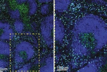
Prof. Ido Amit and Dr. Ziv Shulman of the Department of Immunology, together with their colleagues at Weizmann Institute of Science and in Italy, have developed a method, called NICHE-seq, that combines light-activated fluorescent markers, two-photon microscopy, and single-cell genetic sequencing to infer the cellular and molecular make-up of different immune cell niches.

The study titled "Spatial reconstruction of immune niches by combining photoactivatable reporters and scRNA-seq", published in Science magazine, establishes NICHE-seq as a powerful and reliable method for understanding how spatial location affects immune cell function.

Image (left): Viral infection induces distinct changes in the cellular and molecular composition of specific splenic niches.
Credit: Amit, I, Shulman, Z et al/Science, Dec 2017, eaao4277

Read More about Mapping immune cell niches

Connection is protection

Cell adhesion molecules (CAMs) are proteins located on the surface of the cell. CAMs are involved with intercellular communication. Their role is to help cells stick to each other and to their structural and biochemical support (the extracellular matrix, or ECM).

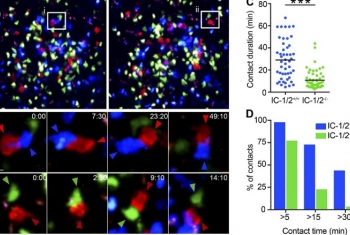
Dr. Ziv Shulman and his research team from the Department of Immunology discovered that a particular cellular adhesion molecule called ICAM is important for selecting the best-suited antibodies for protection against invading pathogens.  

The image (left) shows antibody-forming cells interacting with regulatory T-cells. Blue indicates antibody-forming cells, red indicates T-cells, and green indicates antibody-forming cells that lack adhesion molecule (ICAMs). These cells form shorter contacts with the T-cells.

Image credit: Shulman, Z. et al/Journal of Experimental Medicine, 214 (11): 3435

Migrating from dark zone to light zone

Germinal centers (GCs) are sites within lymph nodes where B cells grow and adapt during a healthy immune response to an infection. GCs separate have two zones, a dark zone and a light zone. As they adapt to suit a particular immune response, maturing B cells migrate from the dark zone to the light zone.

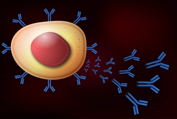
Dr. Ziv Shulman and his team determined that antibody-producing B cells depart from the immune response once they are in the light zone.

The picture (left) shows lymph nodes containing  antibody-forming cells (blue clusters) and cells that depart the immune response (green in D). Cells leave the immune response from the upper side of the lymph node.