Dr. Assaf Gal from the Department of Plant and Environmental Sciences is trying to understand the intracellular mechanisms of minerals formed by unicellular marine algae. With his team, he is focusing on coccolithophores, an abundant group of phytoplankton, which produce coccoliths, mineralized calcite arrays. The formation of the coccolith crystals is exquisitely controlled by the cell. Dr. Gal and his group are striving to understand the fundamentals of the process in order to understand problems related to cell physiology, oceanography, and materials applications.
In a collaboration with Dr. Julia Mahamid at the European Molecular Biology Laboratory, Dr. Gal and Dr. Zohar Eyal, a postdoc working in his lab, have conducted very promising experiments on campus and in Heidelberg, using cryo-electron tomography—an imaging technique used to produce high-resolution three-dimensional views of samples.
Dr. Eyal has established the experimental pipeline in which the calcifying cells are vitrified on a transmission electron microscope grid, and then thinned to 200 nanometers (nm) by a focused ion beam instrument. The final step in this process is acquiring the electron microscope images at different tilt angles. All aspects of this process are now properly functioning in the newly purchased cryo-electron microscopes on campus. Dr. Gal and his team are collecting data from their samples.
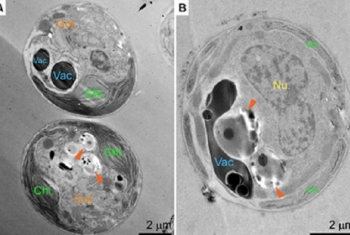
[Image on the left: (A, B) Sections in P. carterae whole cells show both the homogenous lumen of the vacuole that is punctuated with calcium and phosphorus rich (Ca-P) bodies, and the contrasting dense internal organization of the ion-rich compartments (orange arrowheads) that contain particles, spheres, membranes, and more. (Credit: Journal of Structural Biology, 2020)]
