All Activities

Understanding cancer - one cell at a time

The Kleinman Cancer Cell Sorting Facility provides state of the art instrumentation for the study of cancer cells at single cell resolution. Below we highlight some of the recent advances of Weizmann teams using these approaches.

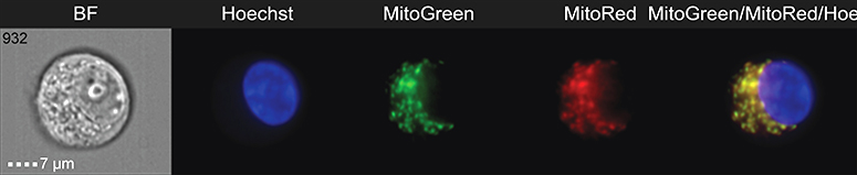
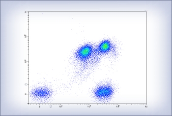
Flow cytometry, the key approach of the Kleinman Cancer Cell Sorting Facility, is a laser based technology in which individual cells are discriminated from each other using surface markers that are 'read' with the help of specific detection reagents - the so-called monoclonal antibodies. This method allows for the analysis of single cell mixtures by passing them in front of a dye-detector at a speed of a thousand cells per second. Moreover, beyond their identification, individual cells can be sorted for subsequent functional assays, as well as molecular analysis, including transcriptome and epigenome profiling.

Revealing the inner cell

By combining the power of high throughput flow-cytometry with a microscope, Image Stream analysis complements the analysis of cell surface markers with critical information on the inside of cells. In the figure shown below from Moshe Oren’s group, distinct localization patterns of mitochondria within cancer cells are depicted.  In blue we see the nucleus of the cells, and in green and red inactive and active mitochondria, which reside within the cytoplasm.

Detecting rare cells

Flow cytometry is a crucial tool for the analysis and diagnosis of blood cell malignancies. Specifically, this technology enabled the Shlush group to detect rare cells that harbor pre-leukemic mutations. The figure (left), made by Amos Tuval, illustrates the cell sorting approach and highlights the discrimination of single cells, which appear as single dots on the two dimensional plot.