Cell Analysis tools
These devices are capable of analyzing single cell samples stained with multiple fluorescent colours. Currently, this unit has two cell analyzers, one with five lasers and another with three lasers. This unit is heavily used by both scientists and students. Due to its popularity, plans are underway to purchase three additional devices in the near future. This will enable the unit to maintain an available device for ad hoc work orders at any given time.
- LSRII-new cell analyzer with five lasers
- LSRII cell analyzer with three lasers

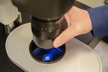
Andor Dragonfly Spinning Disc
The Dragonfly system, with the motorized optical zoom, delivers industry-leading signal to noise and image fidelity. Applications ranging from single molecule to live cell confocal, TIRFM to whole embryo and thick tissue imaging all benefit from the system’s speed and sensitivity.
Cell Sorting
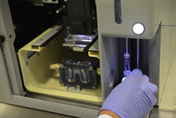
Instruments in this unit serve to analyze and sort cells from chosen populations in a precise manner and deposit them into tubes or plates. This can be done under sterile conditions. The unit currently holds four cell sorters, each with a different number of laser beams and thus a different output. One device also offers analysis with near-UV light.
- FACSAria-FUSION with five lasers. The blue, yellow-green and red can be used either with the violet or the nUV
- FACSAriaIII cell sorter with four lasers
- SORP FACSAriaII cell sorter with five lasers
- FACSAriaII cell sorter with three lasers
Leica TCS SP8 STED with life time imaging module
STED microscopy reaches lateral resolutions below 50 nm, and provides users with the freedom to freely tune the effective focal spot to your needs and achieve super-resolution in x, y and z – online, fast and direct.
CyTOF
This unique device uses mass spectrometry for analysis of single cells by labelling them with stable heavy metal isotopes. The existing Helios device can analyze data with relatively high speed and resolve over 40 metal probes with minimal signal overlap. This unit currently meets scientific needs at the Weizmann Institute, but might expand in the future to include a second device with advanced capabilities.
- Helios Mass Cytometer
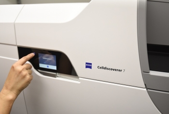
Zeiss Celldiscoverer 7
This system combines the ease-of-use of an automated boxed microscope with the image quality and flexibility of a classic inverted research microscope. Celldiscoverer 7 calibrates itself, detects and focuses on your samples while the optics adjust themselves.
Two-Photon Microscope
Two-photon excitation microscopy allows deep, live imaging of up to a 1mm thickness, and is based in infrared excitation of fluorescent dyes. This method results in a miniscule amount of excitation, in contrast to confocal or wide-field microscopes, and therefore prevents bleaching or other photo-damage to the tissue.
ImageStream
This service is based on the ImageStreamX Flow Imager system, which enables imaging cells in flow. The ImageStream can collect 12 channels of brightfield and fluorescence and quantify staining intensity, pattern, localization, and morphology. As part of the establishment of the Kleinman Cancer Cell Sorting Facility, a second ImageStream device was purchased.
- ImageStreamX Flow Imager systems with 5 lasers (two similar devices)
Lightsheet Z1 – Zeiss
The Lightsheet Z1 by Zeiss is a commercialized lightsheet microscope, this is a fluorescent microscope designed for relatively large live samples for long term time laps, in gentle imaging conditions (relative to conventional confocal microscopy).
The unique Lightsheet Z1 design enables to image samples of three dimensional structures without distortion with close to isotropic resolution in all three axes (X,Y,Z).
The 3D characteristics are also highly valuable for imaging of water based cleared samples of organisms or organs.
UltraMicroscope – II La-Vision
The UltraMicroscope II by La-Vison is a commercialized lightsheet microscope, this is a fluorescent microscope designed for large samples that were usually cleared for imaging using one of the various available protocols.
The ultramicroscope is unique in its ability to image samples clarified in a wide range of refractive index media, from water to organic solvents based protocols.