All Activities

A fly embryo

This is a visualization of transcription dynamics in a live Drosophila fly embryo under Lightsheet microscope.

The nuclei appear in red and the active transcription of the gene snail is detected by the MS2 system resulting in a fluorescent signal. In other words, the green dots inside the nuclei mark the intensity of gene expression.

This tool enables us to study transcription dynamics in a quantitative manner.

Courtesy of Neta Strasser-Rahimi, Benny Shilo lab.

Read More about A fly embryo

Looking at bones

This movie shows a proximal tibial growth plate: red indicates muscles and blood vessels, and green indicates the cartilage.

Courtesy of Sarah Rubin, Eli Zelzer lab

Membranes under microscope

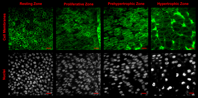
Cell membranes and nuclei of different growth plate zones.

Courtesy of Sarah Rubin, Eli  Zelzer's lab

Read More about Membranes under microscope

Advanced imaging school

Spring school for advanced imaging in biological research was a conference that took place at the Weizmann Institute of Science, on March 15th-16th.

Advanced imaging techniques are becoming increasingly important in life sciences, biological studies and medicine. Technologies span over a wide range, from standard optical microscopy to advanced multiphoton and light sheet imaging, and from magnetic resonance imaging (MRI) to matrix-assisted laser desorption ionization (MALDI).These are very diverse techniques, based on different principles, each isolating a different type of signal or properties. The use of various contrast sources enables aquisition of specific knowledge about an organ, cellular subpopulation or extracellular matrix, providing information about structural features, chemical composition and other characteristics.

During the last decade, resecarhers have combined several methods in order to achieve deeper understanding of live organisms at the cellular and molecular level, for diagnostics and disease modeling - and for the development of novel drugs.

The combination of high spatio-temporal resolution, speed and sensitivity as well as robustness, make today's advanced imaging tools indispensable.

The aim of our school was to maximize the synergy that can be achieved through the use of many research approaches incorporating different disciplines in advanced imaging, as preseted by experts who are world leaders in their fields.

Conference Homepage
Making deep live imaging

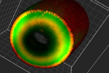
The new two-photon microscope at the de Picciotto-Lesser Cancer Cell Observatory is a powerful tool, which is able to accommodate imaging in human tissue. It serves in the study of a few aspects of cancer, for example, the study of spheroids - 3D cell cultures that are cultivated in vitro to resemble a living tumor.

This system allows deep, live imaging of up to a 1 mm thickness, and is based in infrared excitation of fluorescent dyes. This method results in a miniscule amount of excitation, in contrast to confocal or wide-field microscopes, and therefore prevents bleaching or other photo-damage to the tissue. The excitation process uses a special titanium-sapphire pulsed laser, with very brief peaks of excitation and very high peak power.