Activities
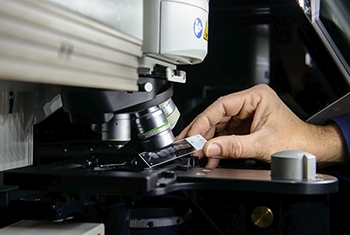
Users can prepare samples in their lab or in the sample preparation lab in the unit. Sample preparation and loading depends on the specific sample and microscope.
- To start working with the microscopes in the unit please contact Sefi Addadi to schedule a meeting. Yoseph.addadi@weizmann.ac.il
- Following the definition of the user's needs, suitable technology and sample preparation, an introduction / test-imaging session will be conducted.
- For System scheduling - click Infernal Service – Advanced light microscopy
- To consult on image analysis using commercial or noncommercial software please contact Ofra Golani ofra.golani@weizmann.ac.il
- For further information check the BioInformatics order page.
- For software license and analysis stations scheduling – click Internal Service – BioImaging Queue Res.

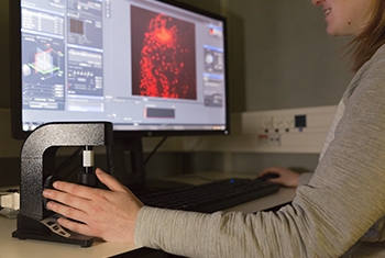
Andor Dragonfly Spinning Disc
The Dragonfly system, with the motorized optical zoom, delivers industry-leading signal to noise and image fidelity. Applications ranging from single molecule to live cell confocal, TIRFM to whole embryo and thick tissue imaging all benefit from the system’s speed and sensitivity.
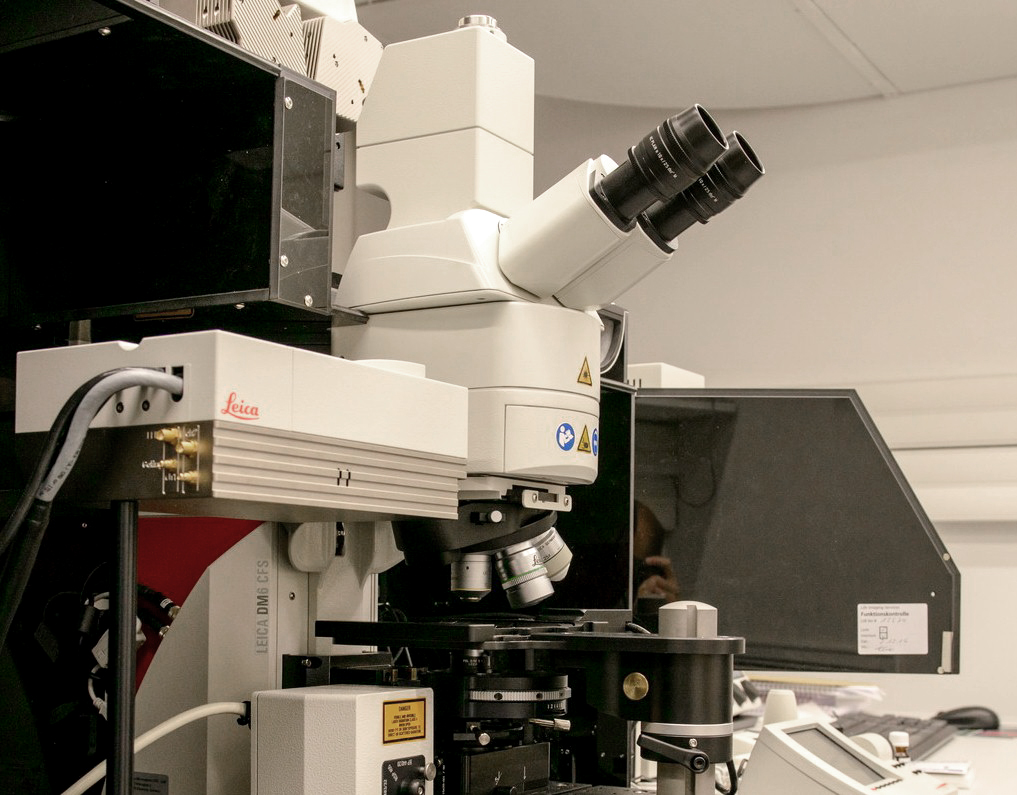
Leica TCS SP8 STED with life time imaging module
STED microscopy reaches lateral resolutions below 50 nm, and provides users with the freedom to freely tune the effective focal spot to your needs and achieve super-resolution in x, y and z – online, fast and direct.
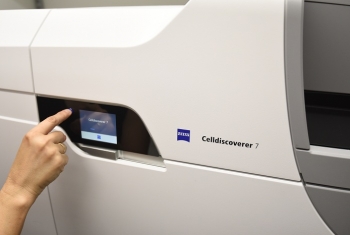
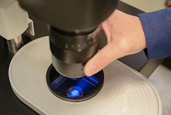
Zeiss Celldiscoverer 7
This system combines the ease-of-use of an automated boxed microscope with the image quality and flexibility of a classic inverted research microscope. Celldiscoverer 7 calibrates itself, detects and focuses on your samples while the optics adjust themselves.
Two-Photon Microscope
Two-photon excitation microscopy allows deep, live imaging of up to a 1mm thickness, and is based in infrared excitation of fluorescent dyes. This method results in a miniscule amount of excitation, in contrast to confocal or wide-field microscopes, and therefore prevents bleaching or other photo-damage to the tissue.
Lightsheet Z1 – Zeiss
The Lightsheet Z1 by Zeiss is a commercialized lightsheet microscope, this is a fluorescent microscope designed for relatively large live samples for long term time laps, in gentle imaging conditions (relative to conventional confocal microscopy).
The unique Lightsheet Z1 design enables to image samples of three dimensional structures without distortion with close to isotropic resolution in all three axes (X,Y,Z).
The 3D characteristics are also highly valuable for imaging of water based cleared samples of organisms or organs.
UltraMicroscope – II La-Vision
The UltraMicroscope II by La-Vison is a commercialized lightsheet microscope, this is a fluorescent microscope designed for large samples that were usually cleared for imaging using one of the various available protocols.
The ultramicroscope is unique in its ability to image samples clarified in a wide range of refractive index media, from water to organic solvents based protocols.