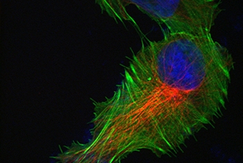
Revealing modification in cell structure
The green, red and blue seen here is the result of an immunostaining technique that reveals modification in cell structure in response to growth factor stimulation. The treatment of a non-cancerous epithelial breast cell line with a growth factor induced motility, leading to the modification and reorganization of the cytoskeleton. This technique, which reveals the presence and position of actin (green), tubulin (red) and the cell nucleus (blue) in non-cancerous cells, enables the visualization of pathways and conformational changes associated with cancer.
The cells were grown on matrix coated glass. The staining was performed through the use of probes for the nucleus (DAPI) and actin (phalloidin),and, for tubulin, with an immunostaining antibody. The images were taken using a spinning disk microscope.
Image curtesy of Lee Roth, Prof. Yosef Yarden lab